Introduction
Tubular adenoma is a rare benign epithelial neoplasm occurring mainly in premenopausal women. The occurrence rate
0.3-1.7% among the benign breast lesions [1]. It is considered
as a variant of fibroadenoma and proposed to have common
histogenesis. However, it has a distinctive histomorphology of
glandular predominance with very sparse intervening stroma
unlike the dense stromal proliferation in fibroadenoma. Previous studies on tubular adenoma have reported its occurrence
along with phyllodes tumor, ductal carcinoma in situ and very
rarely in association with Invasive ductal carcinoma. To the best
of our knowledge this is the first study showing the spectrum of
transformation of fibroadenoma to Pure tubular adenoma in a
same patient. So, we are presenting this case where a 32-year-old female came with complaints of multiple lumps on left side
of breast and found to have Tubular adenoma and fibroadenoma- tubular adenoma mix in different lumps, thereby proving
the common histogenesis.
Case presentation
A 32-year-old lady came with complaints of multiple lumps
in her left breast present for past 1 year. On physical examination 3 lumps were palpable in the upper inner quadrant of left
breast ranging in size from 1 to 3 cm. Ultrasound done revealed
three well circumscribed oval hypoechoic lesions in upper inner
quadrant of left breast measuring 1 cm, 1.4 cm and 2.8 cm. Fine
Needle Aspiration Cytology (FNAC) was requested for the larger
lump and the aspiration smears show low to moderately cellular
smears comprising of ductal epithelial cells along with myoepithelial cells arranged in monolayered sheets and clusters with
individual cells are showing round to oval nucleus with bland
chromatin, moderate cytoplasm and few fibrostromal fragments and scattered bare nuclei seen in a background. So, with
these findings a diagnosis of Fibroadenoma (Category II) (The
Yokohama System for reporting breast cytopathology-2020)
was given. Later on, the two of the lumps with significant size
was excised and sent for histopathological examination.
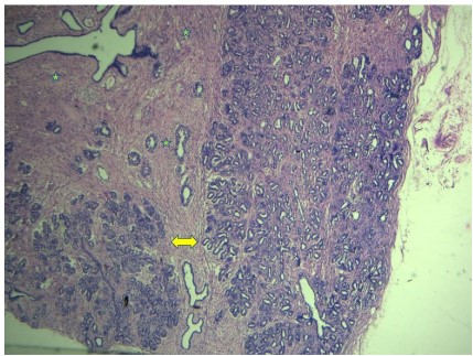
Histopathological examination from the relatively larger
lump show well circumscribed neoplasm consisting of glandular
and stromal proliferation pattern with areas of tubular adenoma (Figure 1). The glands are lined by two layered epithelium.
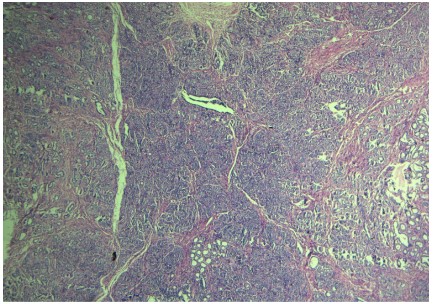
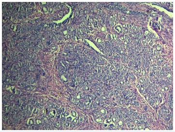
Section examined from smaller lump shows an encapsulated
neoplasm composed of multiple lobules of very closely packed
small sized acini with very minimal intervening stroma (Figures
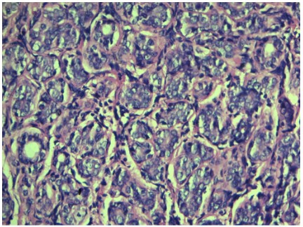
2,3). These acini are lined by luminal epithelial and abluminal
myoepithelial cells (Figure 4). No associated DCIS or invasive
malignancy seen in sections studied. Henceforth, a diagnosis of
Fibroadenoma with areas of tubular adenoma was given to the
larger lump and tubular adenoma was rendered to the smaller
lump.
Discussion
Tubular adenoma is a rare benign epithelial neoplasm accounting for 0.3-1.7% among the benign breast lesions. It is
usually a tumor of young females of premenopausal age group
[1,2] albeit, have also been reported in postmenopausal individuals. Tubular adenomas mostly remain asymptomatic and
rarely associated with breast pain. No alterations seen in overlying skin or nipple [3].
Clinical findings of tubular adenomas are similar to fibroadenoma [4]. They are slow growing and takes 6-12 months before
presentation. Tubular adenomas have occurred in varying sizes
ranging from 1 cm to 7.5 cm, however giant tubular adenomas
of 14 cm size have also been documented in literature [5].
Tubular adenomas are well circumscribed neoplasms with a
homogenous tan yellow cut surface with unremarkable nodularity. Tubular adenomas are difficult to differentiate from fibroadenoma grossly, however the former appears to be softer than
fibroadenomas. Tavassoli et al stated that a nodule to be called
as tubular adenoma it should be at least 1cm or encapsulated
if it is small [4].
Radiologically tubular adenomas and fibroadenomas look
alike. In a study done by Soo et al, where they have analyzed the
imaging findings of 17 patient’s and found that both were well
circumscribed non calcified masses. Similarly, in our case both
the lumps with different histological diagnosis had the same ultrasound findings. Infrequently, imaging findings are worrisome
with microcalcifications, irregular shape, obscure boundary and
uneven echotexture. Even though such features were very rarely reported in tubular adenomas. Radiologists must be aware
that carcinoma can arise from tubular adenoma or present as a
collision in elderly individuals [6].
Table 1: Diagnostic criteria of MIH.
| S.No |
Entityname |
Microscopic findings |
Remarks |
| 1 |
Tubular adenoma |
· Well circumscribed |
Myoepithelial layer intact |
| · small round monomorphic tubules |
| · separated by a barely visible stroma |
| 2 |
Fibroadenoma |
· Well circumscribed |
Myoepithelial layer intact |
| · Both glandular and stromal proliferation |
| 3 |
Tubular adenosis |
· Diffuse infiltrative |
Myoepithelial layer intact |
| · Haphazardly scattered tubules |
| · Abundant fatty or fibrous stroma |
| 4 |
Microglandular adenosis |
· Poorly circumscribed |
Myoepithelial layer absent, however basement membrane intact |
| · Small uniform glands |
| · Open lumina |
| · Embedded in a adipocytic /fibrous stroma |
| · Glands lined by cuboidal cells with uniform heightand pale cytoplasm |
5 |
Sclerosing adenosis |
Not sharply circumscribed, but localized Lobulocentric pattern |
Myoepithelial layer intact- usually spindled |
| · Compressed glands |
| 6 |
Radial scar |
· Lobulocentric pattern |
Myoepithelial layer intact |
| · Central fibrosis |
| · Tubules are not monomorphic |
| · Abundant fibrous stroma |
| 7 |
Phyllodes tumorbenign |
· Well circumscribed |
Myoepithelial layer intact |
| · Glandular and stromal proliferation |
| · Leaf like architecture |
| 8 |
Tubular carcinoma |
· Stellate infiltrative |
Myoepithelial layer and basement membrane absent |
| · Angulated glands |
| · Wide open lumina |
| · Embedded in a desmoplastic stroma |
Cytological findings of tubular adenoma are very sparse.
Three-dimensional ball like clusters and small tubular structures
are reported as the predominant findings in Cytology. Tubular
adenosis and tubular carcinoma are the other differentials to
be considered when tubular structures are seen in cytology [7].
Our case no FNAC was attempted from the tubular adenoma
lump and the FNA done from fibroadenoma with tubular adenosis didn’t reveal any tubular structures as one can except.
Histopathological examination of tubular adenomas will
show well circumscribed or well encapsulated neoplasm comprising of small round uniform tubules lined by luminal epithelial and abluminal basal cells. The acini are usually separated by
a sparse, barely visible stroma [2]. Our case had similar type of
histological features.
Both benign and malignant neoplasms can often create diagnostic dilemmas with tubular adenomas and a number of
lesions enter into the differential’s like fibroadenoma, microgranular adenosis, sclerosing adenosis, radial scar, tubular carcinoma etc., [8]. So, this has to be correlated with clinical and
radiological findings and can be supported by Immunohistochemistry (IHC) if needed. The main diagnostic features of each
entity are given in table 1.
IHC is not routinely required for diagnosing tubular adenomas. At times when there is a confusion with tubular carcinoma
or co-existing carcinoma, a battery of myoepithelial markers
like Smooth Muscle Actin (SMA), p63, CK5/6, calponin can be
thrown to find the intactness of myoepithelial cells and ruling
out carcinomas [9]. In our case the morphological features were very obvious for tubular adenoma and hence we didn’t require
Immunohistochemical support.
With regard to the histogenesis of tubular adenoma. Few
authors consider it is an extreme variant of fibroadenomas and
backing a common histogenesis for both [10]. Study by Maiorano et al., have revealed that the histomorphology and immunohistochemical features of both these entities are similar in few
areas [9]. In our study we found one lump with fibroadenoma
admixed with tubular adenoma like areas and another lump
in same patient with pure tubular adenoma. This makes us to
think in line with the above theory and this is viable evidence
supporting the notion of common histogenesis.
The main stay of management is surgical excision. Complete
excision is often curative. Core biopsy can be done before complete excision for correct diagnosis [11].
Conclusion
Tubular adenomas are rare benign epithelial neoplasms
sharing a common histogenesis with fibroadenomas and mimic
fibroadenomas multidimensionally and our case is one other
evidence for that. Tubular adenomas can occur alone or as an
intimate admixture with fibroadenoma or phyllodes tumor.
Since there are a number of great mimickers for this entity adding to further confusions and diagnostic dilemmas, awareness
of this entity is essential because it is totally benign and curative
by surgical excision.
References
- Tavassoli FA, Devilee P. World Health Organization Classification
of Tumours. Pathology and Genetics of Tumours of the Breast
and Female Genital Organs. IARC Press: Lyon. 2003.
- Lakhani SR, Ellis IO, Schnitt SJ, Tan PH, Van de Vijver MJ, eds.
WHO Classification of Tumours of the Breast. 4th ed. Lyon,
France: IARC; 2012.
- Hertel BF, Zaloudek C, Kempson RL. Breast adenomas. Cancer.
1976; 37: 2891-2905
- Tavassoli FA. Benign lesions. In: Pathology of the Breast. 2nd ed.
Stamford, Conn: Appleton& Lange. 1999; 115-204.
- Düşünceli F, Manukyan MN, Midi A, Deveci U, Yener N. Giant
tubular adenoma of the breast: A rare entity. Breast J. 2012; 18:
79-80.
- Soo MS, Dash N, Bentley R, Lee LH, Nathan G. Tubular adenomas
of the breast: imaging findings with histologic correlation. Am J
Roentgenol. 2000; 174: 757-61.
- Shet TM, Rege JD. Aspiration cytology of tubular adenomas of
the breast. An analysis of eight cases. Acta Cytol. 1998; 42: 657-662.
- Efared B, Sidibé IS, Abdoulaziz S, Hammas N, Chbani L, et al.
Tubular Adenoma of the Breast: A Clinicopathologic Study of a
Series of 9 Cases. Clin Med Insights Pathol. 2018; 5: 11.
- Maiorano E, Albrizio M. Tubular adenoma of the breast: an immunohistochemical study of ten cases. Pathol Res Pract. 1995;
191: 1222-123.
- Le gal y. Adenomas of the breast: relationship of adenofibromas
to pregnancy and lactation. Am Surg. 1961; 27: 14-22.
- Zuhair AR, Maron AR. Tubular adenoma of the breast: A case
report. Case Reports Clin Med. 2014; 3: 323-6.


